Key Takeaways
- Gout is caused by monosodium urate crystals; pseudogout (CPPD) by calcium pyrophosphate crystals. They are different diseases. [1]
- Gout classically strikes the big toe; pseudogout most often affects the knee, followed by the wrist. [1][2]
- Serum urate is usually high in gout and usually normal in pseudogout, though the two can coexist. [1][3]
- Only crystal analysis of joint fluid identifies which crystal is present and definitively tells them apart. [4]
- Pseudogout has no urate-lowering drug; acute attacks are treated with NSAIDs, corticosteroids, or colchicine. [2]
- Pseudogout should prompt a check for associated conditions such as hyperparathyroidism, hemochromatosis, low magnesium, and thyroid disease. [1][2]
Two Different Crystals
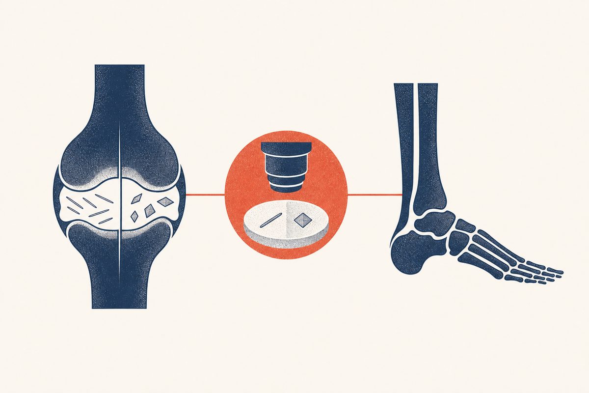
The two diseases are close mimics because both produce a sudden, hot, swollen, painful joint. The difference is at the microscopic level. Gout forms needle-shaped monosodium urate crystals when serum urate is too high. Pseudogout, or calcium pyrophosphate deposition (CPPD) disease, forms rhomboid, weakly positively birefringent calcium pyrophosphate dihydrate crystals in cartilage, often without any urate problem at all.[1]
Because treatments diverge, it matters which crystal is present. Gout is managed toward a low serum urate target with drugs such as allopurinol; pseudogout has no urate-lowering medicine and is managed with anti-inflammatory treatment of attacks and attention to any associated metabolic condition.[2][3]
| Crystal feature | Gout | Pseudogout (CPPD) |
|---|---|---|
| Composition | Monosodium urate | Calcium pyrophosphate dihydrate |
| Shape | Needle-shaped | Rhomboid (rod or parallelepiped) |
| Polarized light | Strongly negatively birefringent | Weakly positively birefringent |
| Where it forms | In and around joints when urate is high | In cartilage (chondrocalcinosis) |
Which Joints Each Affects
The first metatarsophalangeal joint (the big toe) is the classic gout joint, the attack called podagra. Gout also affects the midfoot, ankle, and knee.[3] Pseudogout is most common in the knee, and its other favored sites are the wrist, shoulder, and the small joints of the hand, a pattern that differs from typical gout.[1][2]
Joint pattern is a helpful clue but not proof. A knee flare in an older adult is more likely CPPD, while a first big-toe flare in a middle-aged man with high urate is more likely gout. The diagnosis is confirmed by crystals, not by location alone.[1]
Serum Urate: High vs Normal
In gout, serum urate is typically above the 6.8 mg/dL saturation point, the higher it is and the longer it stays there, the higher the gout risk.[3] In pseudogout, the serum urate is usually normal, because calcium pyrophosphate, not urate, is the problem.[1] This is a practical clue at the bedside, but it is not diagnostic: urate can be normal during a gout flare, and a person with CPPD can also have high urate. The two conditions can coexist, particularly in older adults.[1][3]
Who Gets Which
Gout is more common in men and its risk rises from middle age, tied to hyperuricemia, obesity, kidney disease, medications such as diuretics, alcohol, and diet.[5] Pseudogout is strongly age-related and becomes common in people over 60, affecting men and women more equally as age climbs. Both can appear together in older adults, which is why a single hot joint in an older person still needs a careful look.[1][2]
| Clinical clue | Points toward gout | Points toward pseudogout |
|---|---|---|
| Age and sex | Middle-aged men | Older adults (60+), men and women |
| Joint | First toe, midfoot, ankle | Knee, wrist, hand |
| Serum urate | Usually elevated | Usually normal |
| Recent trigger | Alcohol, purine-rich meal, dehydration | Illness, surgery, trauma |
| Imaging | Urate deposits, bone erosions | Chondrocalcinosis on X-ray |
How the Diagnosis Is Made
The definitive test is joint-fluid analysis: removing a small sample of fluid with a needle and examining it under a polarized-light microscope. Gout shows strongly negatively birefringent, needle-shaped urate crystals; pseudogout shows weakly positively birefringent, rhomboid calcium pyrophosphate crystals.[1][4] This single step both confirms the crystal disease and rules out septic arthritis, which can look identical and can destroy a joint within days.[4]
Imaging adds support. In gout, ultrasound can show a double-contour sign and dual-energy CT can show urate deposits; in CPPD, X-rays classically show calcium lines in cartilage (chondrocalcinosis). These findings support but do not replace the crystal confirmation when the diagnosis is uncertain.[1][4]
What Causes Pseudogout
CPPD is mostly a consequence of aging cartilage, but in younger people or in widespread or recurrent disease it can signal an underlying metabolic condition. The associations to consider include hyperparathyroidism, hemochromatosis, hypomagnesemia, hypophosphatasia, and sometimes thyroid disease.[1][2]
Unlike gout, pseudogout is not caused by diet or by a high uric acid level, and reducing purines or urate does not treat it. Attacks can be triggered by illness, surgery, trauma, or a medical stress, which is a different trigger profile from the overnight, diet-and-alcohol-adjacent picture of gout.[1]
How Treatment Differs
| Gout | Pseudogout (CPPD) | |
|---|---|---|
| Crystal | Monosodium urate (needle-shaped) | Calcium pyrophosphate (rhomboid) |
| Serum urate | Usually high | Usually normal |
| Classic joint | First toe | Knee, wrist |
| Acute treatment | Colchicine, NSAID, or steroid | NSAID, steroid (often joint injection), or colchicine |
| Long-term strategy | Urate-lowering drug (allopurinol) | No urate-lowering drug; manage recurrences and associated conditions |
Acute attacks of pseudogout are treated very much like gout, with an NSAID, an intra-articular or systemic corticosteroid, or colchicine; joint aspiration itself, by removing fluid and crystals, also relieves a knee attack.[2] The major divergence is long-term: gout has a disease-modifying strategy in urate-lowering therapy, while CPPD does not, so recurrent pseudogout is managed with occasional prophylaxis and treatment of any underlying metabolic disorder, not with allopurinol.[1][2][3]
Can You Have Both?
Yes. Gout and CPPD can coexist in the same person and even in the same joint, most often in older adults. When both crystals are present, the clinician manages the urate target for the gout component while treating flares and any associated disorder for the CPPD component. This is another reason crystal identification, rather than guessing from urate alone, guides care.[1]
When a Hot Joint Needs Urgent Care
Seek same-day emergency evaluation for fever or chills with a hot joint, rapidly spreading redness, a wound near the joint, inability to bear weight, or severe illness. The most important distinction in an acutely hot joint is not gout versus pseudogout, it is either versus septic arthritis, which requires prompt joint-fluid analysis and antibiotics.[4]
See our guides on gout and gout attack treatment for the fuller picture of urate-driven disease.
Frequently Asked Questions
No. They look alike but involve different crystals. Gout is urate crystals from high uric acid; pseudogout is calcium pyrophosphate crystals, usually with normal uric acid.[1]
Acute attacks are treated with an NSAID, a corticosteroid (often injected into the joint), or colchicine. There is no urate-lowering drug for pseudogout, and allopurinol does not treat it.[2]
Similar to gout: sudden severe pain, swelling, warmth, and stiffness, often in the knee. Attacks can follow illness, surgery, or injury.[1]
No. Unlike gout, dietary purines and alcohol are not the cause of pseudogout. It is a disorder of calcium pyrophosphate deposition in cartilage.[1]
Yes, especially in older adults. Both crystals can be present, and management addresses the urate for gout plus flare treatment and any associated condition for pseudogout.[1]
Yes. Calcium pyrophosphate deposition rises steeply after age 60 and is rare in people under 50, which is a different age profile from gout, which starts earlier in many men.[1]
Tophi are a feature of urate (gout), not calcium pyrophosphate disease. CPPD causes cartilage calcification and joint damage rather than urate tophi.[1]
References
- Rosenthal AK, Ryan LM. Calcium Pyrophosphate Deposition Disease. New England Journal of Medicine. 2016;374(26):2575-2584. doi:10.1056/NEJMra1511117. PMID: 27355536. https://pubmed.ncbi.nlm.nih.gov/27355536/
- Zhang W, Doherty M, Pascual E, et al. EULAR recommendations for calcium pyrophosphate deposition. Part II: management. Annals of the Rheumatic Diseases. 2011;70(4):571-575. doi:10.1136/ard.2010.139360. PMID: 21257614. https://pubmed.ncbi.nlm.nih.gov/21257614/
- FitzGerald JD, Dalbeth N, Mikuls T, et al. 2020 American College of Rheumatology Guideline for the Management of Gout. Arthritis Care & Research. 2020;72(6):744-760. doi:10.1002/acr.24180. https://pmc.ncbi.nlm.nih.gov/articles/PMC10563586/
- Newberry SJ, FitzGerald JD, Motala A, et al. Diagnosis of Gout: A Systematic Review in Support of an American College of Physicians Clinical Practice Guideline. Annals of Internal Medicine. 2017;166(1):27-36. doi:10.7326/M16-0462. PMID: 27802505. https://pubmed.ncbi.nlm.nih.gov/27802505/
- Kuo CF, Grainge MJ, Zhang W, Doherty M. Global epidemiology of gout: prevalence, incidence and risk factors. Nature Reviews Rheumatology. 2015;11(11):649-662. doi:10.1038/nrrheum.2015.91. PMID: 26150127. https://pubmed.ncbi.nlm.nih.gov/26150127/